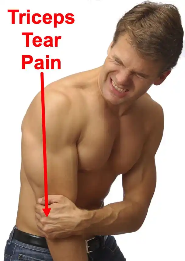
Distal Triceps Rupture
What is a triceps tendon tear?
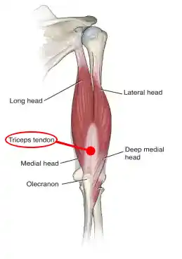
The triceps is the large muscle at the back of the upper arm. The triceps consists of three separate muscles that form a single combined tendon which attaches to the back of the elbow. Tears may be partial or complete. A complete / full tear (rupture) typically occur when a sudden large load is applied to the triceps (eg. falls, weightlifting). Some complete ruptures are associated with pre-existing conditions (tendonitis), injuries (partial tears) or drug treatment (oral steroid use, local steroid injections). The triceps normally straightens the arm; when a triceps tendon tear occurs, there is weakness with elbow extension. Men who participate in sports, and who are ages 30-50 are particularly at risk.

Causes of a Distal Triceps Tear
The most common causes of a distal triceps tear include:
- Mountain Biking
- Fall causing sudden forceful bending of the elbow
- Heavy lifting
- Sports injuries
The risk for a distal triceps tear is greater in the presence of the following:
- Pre-existing conditions such as tendonitis
- Drug treatment such as use of steroids
- Autoimmune diseases
Symptoms of a Distal Triceps Tear
Symptoms of a distal triceps tendon rupture may include:
- A popping-out sensation felt at the back of the elbow.
- A cracking or snapping sound at the time of injury.
- Severe pain initially that gradually recedes.
- Triceps muscle cramping.
- Swelling and tenderness in the elbow.
- Triceps swelling or slight bulging at the back of the arm.
- Weakness in the arm.
- Difficulty with elbow range of motion.
Diagnosis of a Distal Triceps Tear
A distal triceps tear is diagnosed typically by a thorough physical evaluation. You may be asked to perform triceps extension tests to rule out whether the tendon tear is partial or complete. Your medical history will be discussed. Your provider may order imaging studies to confirm the diagnosis such as an X- ray, ultrasound or MRI scan to look for abnormalities. MRI is considered the gold standard technique for diagnosis because it can demonstrate soft-tissue inflammatory changes of the tendon, as well as torn and retracted tendon fibres.
Treatment of a Distal Triceps Tear
Distal triceps tear management includes the following:
Non-surgical treatment:
Conservative treatment methods are used if you have suffered a partial tendon tear. Most partial triceps tendon injuries will heal over 8-12 weeks. The treatment options incude:
- Rest or immobilisation of the arm or elbow with a sling or splint.
- Ice pack or hot water bag application to reduce inflammation.
- Anti-inflammatory medicines to reduce swelling.
- Pain relievers to reduce pain.
- Physical therapy and rehabilitation.
Surgical treatment:
A complete tear of the triceps tendon will require surgery. Surgery involves reattaching the tendon to the ulna in order to avoid permanent elbow extension weakness and muscle atrophy.
Surgery would be planned typically within 1-2 weeks of diagnosis of a complete complex rupture. Triceps tendon repair is done by reattaching the tendon to the bone using drill holes or bone anchors. Following surgery, a splint should be used to immobilise the elbow for one to two weeks, and later replaced with a removable brace.
Physical therapy may be started gradually, depending on the healing process. You may resume your normal activities within 3-4 months.
Definition
A triceps tendon rupture / tear occurs when the tendon tears or avulses from its insertion point on the olecranon, resulting in difficulty or inability to extend the elbow against gravity or resistance. Complete ruptures involving all three heads of the triceps tendon (long, lateral, and medial) typically require surgical intervention. Partial tears may be tolerated in patients with low functional demands.
Triceps tendon ruptures are usually caused by a sudden, forceful contraction of the triceps muscle against resistance or direct trauma to the elbow. The injury can be associated with other conditions such as olecranon bursitis or systemic diseases like rheumatoid arthritis.
Natural History
If untreated, complete ruptures can lead to significant weakness in elbow extension and functional impairment. Partial tears may heal with conservative treatment in patients with lower functional demands, but they might also lead to chronic pain and weakness if not properly managed.
Patient History and Physical Findings
Patients typically present with:
- Sudden onset of pain at the back of the elbow.
- A popping or snapping sensation at the time of injury.
- Swelling and bruising around the elbow.
- Palpable gap or defect above the olecranon.
- Difficulty or inability to extend the elbow against gravity or resistance.
Imaging and Other Diagnostic Studies
- X-Rays: May show an avulsion fracture of the olecranon.
- MRI: Useful for confirming the diagnosis, assessing the extent of the tear, and identifying any associated injuries. MRI often demonstrates a bipartite insertion between the deep and superficial components of the triceps tendon into the olecranon.
Differential Diagnosis
- Olecranon bursitis
- Distal humerus fracture
- Elbow dislocation
- Lateral epicondylitis
- Medial epicondylitis
Nonoperative Management
- Indications: Partial tears in patients with low functional demands.
- Treatment: Immobilization in a splint or brace followed by a gradual rehabilitation program focusing on restoring range of motion and strengthening the triceps muscle.
Surgical Management
Indications: Complete ruptures or partial tears in patients with high functional demands.
Procedure: Reattachment of the triceps tendon to the olecranon using suture anchors, transosseous sutures, or other fixation methods. Postoperative rehabilitation includes immobilization followed by gradual range of motion and strengthening exercises.
Postoperative Care
- Immobilization: The elbow is typically immobilized in a splint or brace for several weeks to allow for initial healing.
- Rehabilitation: Gradual range of motion exercises begin after immobilization, followed by progressive strengthening exercises to restore full function.
Outcomes
With appropriate treatment, most patients can expect to regain full strength and function in the affected arm. Early diagnosis and intervention are critical to achieving the best outcomes and preventing long-term disability.
Rehabilitation
A graduated rehabilitation program over 12 weeks focuses on:
- Immobilization for 1-2 weeks
- Gradual range of motion from weeks 2-6 using a brace
- Isometric and resistance exercises from weeks 6-12
Frequently Asked Questions
Can a ruptured triceps tendon heal without surgery?
Partial tears may heal conservatively, but complete tears require surgical repair for optimal function.
How long does it take to recover from triceps tendon surgery?
Full recovery can take 4-6 months with rehabilitation.
What is the success rate of triceps tendon surgery?
Surgical repair generally restores good function, but strength may not return to 100% of the uninjured side.