Understanding Radiocapitellar Plica
An Educational Guide to Radiocapitellar Plica Syndrome
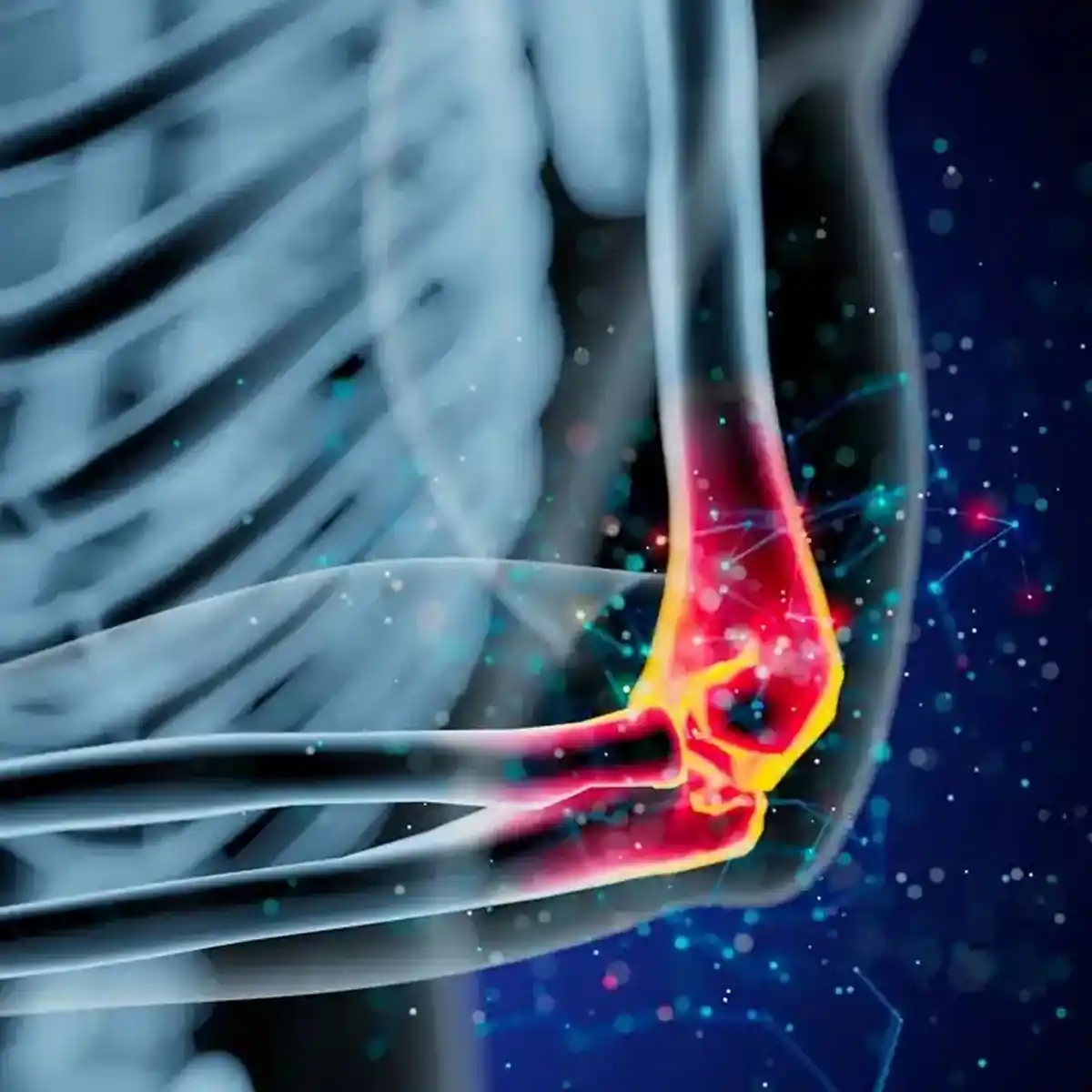
Radiocapitellar plica syndrome is a condition of the elbow that can cause pain and mechanical symptoms like snapping or popping. It involves a "plica," which is a normal fold of the synovial tissue that lines the inside of the elbow joint. While these folds are present in many people and cause no issues, a plica can become a source of pain if it gets thickened, inflamed, or pinched between the bones of the joint during movement. This condition specifically affects the radiocapitellar joint, which is on the outer (lateral) side of the elbow. Understanding this diagnosis is important because its symptoms can mimic other common elbow problems.
Detailed Information on Radiocapitellar Plica
All About Radiocapitellar Plica Syndrome
A radiocapitellar plica is a fold of synovial tissue located in the outer part of the elbow joint, between the radius and the humerus bones. In most individuals, this tissue is thin and flexible, allowing for smooth motion. However, this plica can become symptomatic if it undergoes changes, turning into a larger, inflamed, or scarred (fibrotic) structure. When this happens, the thickened fold can get caught or impinged during certain elbow movements, leading to pain and other symptoms. This condition is often considered a diagnosis of exclusion, meaning other potential causes of lateral elbow pain are ruled out first.
Causes and Risk Factors
The transition of a normal, asymptomatic plica into a problematic one can be triggered by several factors.
- Repetitive Overload: Repetitive motions, such as those in certain sports or occupational tasks, can cause microtrauma to the synovial fold, leading to irritation and thickening over time.
- Direct Injury: A direct blow to the elbow or a fall can cause inflammation within the joint, which may lead to the plica becoming symptomatic.
- Joint Irritation: Any condition that causes ongoing inflammation in the elbow joint can potentially lead to the thickening and fibrosis of a synovial fold.
Symptoms
The symptoms of radiocapitellar plica syndrome are primarily related to mechanical impingement.
- Lateral Elbow Pain: Pain is typically felt on the outer side of the elbow, often in the posterolateral region (toward the back and outer side).
- Painful Snapping or Popping: A distinct snapping or popping sensation may be felt during elbow movement, particularly with a combination of bending the elbow and rotating the forearm.
- Locking or Pain with Extension: Some individuals experience a feeling of the elbow locking or have pain when trying to fully straighten the arm (terminal extension).
- Worsening with Movement: Pain is often aggravated by activities that involve extremes of elbow motion.
Diagnosis
Diagnosing radiocapitellar plica syndrome involves a careful clinical evaluation, often supported by imaging to rule out other conditions.
- Physical Examination: A clinician will assess the elbow for areas of tenderness, which is often found over the posterolateral radiocapitellar joint. Provocative maneuvers, such as the flexion-pronation test (bending the elbow while turning the palm down), may be used to reproduce the painful snapping. Pain at terminal extension is also a common finding.
- X-rays: X-rays are usually normal in cases of isolated plica syndrome but are useful for ruling out other issues like arthritis or loose bodies in the joint.
- Ultrasound: Dynamic ultrasound can be a valuable tool for evaluating lateral elbow pain and can sometimes visualize the plica moving or snapping during motion.
- MRI: An MRI can show the synovial folds and any associated cartilage changes. However, it can be difficult to distinguish a symptomatic plica from a normal one based on imaging alone, as their thickness can overlap.
- Arthroscopy: In some cases, arthroscopy (a minimally invasive surgical procedure) may be used to directly visualize the inside of the joint, confirm that the plica is impinging, and rule out any other intra-articular causes of pain.
Frequently Asked Questions
What is the difference between a plica and a tendon?
A tendon is a tough, fibrous cord that connects muscle to bone, designed to transmit the force of muscle contractions to create movement. A plica, on the other hand, is a fold in the synovium, which is the soft, thin membrane that lines the inside of a joint. While both can cause pain, tendon problems (like tennis elbow) are typically painful when the associated muscle is used against resistance, whereas plica syndrome pain is more often related to the mechanical pinching of the fold during specific joint movements.
Can an MRI definitively diagnose radiocapitellar plica syndrome?
While an MRI is excellent for visualizing soft tissues like a plica, it cannot always definitively diagnose the syndrome. Research has shown that synovial folds are a normal finding in the elbow, and there is often an overlap in the thickness of plicae in people with and without symptoms. Therefore, while an MRI can identify a plica and help rule out other problems like cartilage damage or loose bodies, the diagnosis of plica syndrome is ultimately a clinical one, based on the combination of your symptoms, physical exam findings, and imaging results.
Is painful snapping in the elbow always caused by a plica?
No, painful snapping in the elbow can have several causes. Besides a radiocapitellar plica, snapping can be caused by an unstable ulnar nerve ("funny bone" nerve), a snapping triceps tendon, or a loose piece of cartilage or bone within the joint. Each of these conditions has a slightly different character and location of snapping. A thorough examination by a clinician is necessary to perform specific tests that help differentiate between these possible causes and arrive at an accurate diagnosis for your elbow snapping.